Urolithiasis in rabbits (urolithiasis, urinary calculi) is disease characterised by formation of calculi in organs of the urinary system.
Pathogenesis and aetiology
- Urinary obstruction causes dilation proximal to the obstruction (ureteric dilation / bladder distension). Subsequent changes depend on site, degree, and duration of obstruction; acute kidney injury may develop;
- The most common cause of urinary obstruction in rabbits is hypercalciuria with formation of uroliths and “sand” blocking the urethra. Almost all dietary calcium is absorbed and excreted mainly by the kidneys; roughly 45–60% in rabbits compared with less than 2% in many other mammals;
- Rabbit diets are often high in calcium, yet not all rabbits develop hypercalciuria;
- The aetiology of hypercalciuria and urolithiasis is not fully elucidated; disease is more common in overweight, sedentary animals fed predominantly alfalfa;
- Oligodipsia leading to concentrated urine and factors impairing complete bladder emptying (limited exercise, obesity, cystitis, neoplasia, neuromuscular disease) may contribute. With oliguria calcium crystals precipitate in the bladder and settle as thick “sand” that does not mix with urine or pass during voiding, forming uroliths;
- Calculi may also form in the renal pelvis or ureters.
It is most often seen in rabbits aged 3–5 years.
Signs of urolithiasis in rabbits
- Pollakiuria or stranguria;
- Before complete obstruction owners may note thick paste-like beige-brown urine; sometimes it is so thick it is mistaken for diarrhoea;
- Disease may be asymptomatic despite heavy sediment in the bladder; urine may be clear or slightly cloudy;
- Urine scald dermatitis;
- Macroscopic haematuria;
- Signs of uraemia when the urethra is obstructed: anorexia, weight loss, lethargy, bruxism, tenesmus, hunched posture.
Physical examination
- Distended, tense bladder;
- Urolith palpable (absence on palpation does not rule out urolithiasis). Usually one large stone is palpable; with crystalluria the bladder may feel soft and crumbly;
- Expression of the bladder may yield thick beige-brown urine;
- Palpably enlarged kidneys with stones and hydronephrosis (uncommon);
- Signs of severe uraemia: dehydration, weakness, hypothermia, bradycardia or tachycardia, shallow breathing, stupor or coma, seizures, severe hyperkalaemia.
Risk factors
- Inadequate water intake (dirty bottles, unpalatable water, change of water source, restricted access);
- Neuromuscular disease or pain that discourages urination (musculoskeletal disease, dental disease, abscesses, etc.);
- Inadequately clean litter or cage leading rabbits to avoid urinating for abnormally long periods;
- Obesity;
- Lack of exercise;
- Diet based exclusively on alfalfa;
- Kidney failure;
- Calcium or vitamin–mineral supplements.
Laboratory tests
Blood results are often near normal.
Possible changes:
- Leukocytosis with urinary tract infection;
- Complete obstruction may cause post-renal azotaemia (elevated urea, creatinine, potassium, or phosphorus).
Urinalysis may show calcium oxalate or carbonate crystals — a common finding in rabbits and not necessarily disease. Pyuria, haematuria, or proteinuria may indicate urinary tract disease.
Before antibiotics, obtain urine culture by cystocentesis or bladder wall sample intraoperatively.
Imaging
Abdominal radiography
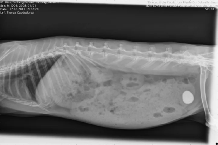
- Calcium oxalate uroliths are radiopaque and usually visible on survey radiographs. Uroliths must be differentiated from bladder sediment. Ultrasound and palpation help distinguish single stones from amorphous sand;
- Relatively large stones may lodge in the urethra causing only partial obstruction;
- Stones may be found in kidneys or ureters.
Abdominal ultrasound
Ultrasound is sensitive for stone location and for assessing anatomical changes in kidneys or ureters (hydronephrosis, ureteritis, perforation, etc.).
Treatment of urolithiasis in rabbits
- Complete obstruction is an emergency and may be life-threatening; treatment must begin immediately;
- Partial obstruction is not always an emergency but these patients are at risk for complete blockage or irreversible urinary tract damage and need timely treatment;
- Hospitalisation until spontaneous urination returns;
- Surgery is sometimes required;
- Long-term outlook depends on cause of obstruction and degree of bladder wall or renal parenchymal damage;
- NSAIDs (meloxicam 0.3–0.6 mg/kg), antibiotics per culture results, intravenous fluids to manage uraemia.
If you are unsure about your pet’s diagnosis and want a second opinion, you can request a veterinary consultation via messengers, phone, or the form on this page.