Spontaneous hyperadrenocorticism is a disorder of excessive secretion by the adrenal cortex of oestrogens, androgens, and related compounds. Clinical signs may reflect systemic effects of circulating sex steroids and local mass effects of tumours. Serum cortisol is often not markedly elevated in most ferrets with adrenal disease — unlike classical hyperadrenocorticism in dogs.
Pathophysiology
Adrenal disease in ferrets is driven by excessive secretion of sex steroids from adrenal cortical hyperplasia, adenoma, or carcinoma. Unilateral adrenal involvement is somewhat more common than bilateral.
Some classic hyperadrenocorticism categories are poorly documented in ferrets: ACTH-secreting pituitary tumours and iatrogenic hyperadrenocorticism from excessive exogenous glucocorticoids.
Systems affected
Adrenal disease in ferrets is multisystem; skin and reproductive tract predominate. Bone marrow suppression may occur.
Involvement varies — some animals show mainly one system, others several equally.
Epidemiology
Hyperadrenocorticism is among the most common diseases of pet ferrets, affecting up to ~70%. Over 95% of ferrets with bilateral symmetric progressive alopecia have adrenal disease.
It is seen mainly in surgically neutered animals; sex incidence is similar; females often present because of obvious vulvar swelling. Typical onset is 3–4 years (literature range ~1–7 years).
Clinical signs
- Severity varies with duration and excess sex steroid levels; mass effects and catabolic tumour effects may contribute;
- Alopecia is the most common sign — sudden progressive hair loss or seasonal onset that regresses then recurs; usually bilateral symmetric loss beginning at tail base progressing cranially; diffuse thinning or shoulder bald patches may occur; skin often looks normal but may seem thickened; severe cases may become nearly hairless;
- ~30% scratch; secondary pyoderma may occur;
- swollen vulva in neutered females is very common;
- stranguria from paraurethral/urogenital cysts, abscesses, or prostatic hyperplasia (males) — common and potentially life-threatening; large masses may be palpable;
- sexual aggression or return of sexual behaviour in neutered animals;
- thin skin, muscle wasting, abdominal enlargement in chronic cases;
- mammary hypertrophy (sometimes);
- anaemia, polydipsia, polyuria (rare);
- enlarged adrenal palpable (sometimes);
- splenomegaly — common, often incidental.
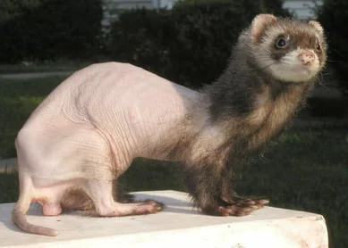
Alopecia in adrenal disease. Photo from AllFerrets
Risk factors
Evidence links adrenal disease to early surgical neutering; gonads and adrenals share urogenital ridge embryology and some gonadal-like cells may persist in adrenals — gonadotroph stimulation could hypertrophy steroid-secreting cells.
Differential diagnoses
- for hair loss and pruritus: seasonal alopecia, pyoderma, mast cell tumour, cutaneous lymphoma;
- for swollen vulva in neutered females: ovarian remnant;
- for stranguria: cystitis, urolithiasis.
Laboratory tests
Bloodwork is often normal. Chronic cases may show non-regenerative anaemia, leukopenia, or thrombocytopenia from oestrogen excess.
Leukocytosis occurs with abscessed urogenital cysts or uterine stump pyometra.
Hypoglycaemia may reflect concurrent insulinoma — common in this age group.
Urinalysis may show low specific gravity in polyuric animals (rare).
Plasma oestradiol, androstenedione, and 17-hydroxyprogesterone together are the most diagnostically useful markers. Concentrations often drop markedly after removal of affected gland(s) with resolution of signs.
Elevated oestrogens in males are significant; in females oestrogen may also rise from ovarian remnants.
Elevated cortisol is uncommon.
Unlike dogs, ACTH stimulation and low-dose dexamethasone tests are not meaningful for adrenal disease diagnosis in ferrets.
Imaging
Radiographs may show adrenal enlargement.
Structures around the bladder may be visible with prostatomegaly or urogenital cysts.
Ultrasound is most useful to visualise enlarged adrenal glands. Normal width × length ~2–3.7 mm × 4–8 mm. Diseased glands often have widened poles (>3.9 mm), rounded shape, abnormal echogenicity, or mineralisation. Some functional disease exists with initially normal ultrasound — screening abdominal ultrasound is advised in older ferrets.
Diagnostic procedures
100 IU human chorionic gonadotropin (hCG) IM once, no sooner than 2 weeks after vulvar swelling appears. If swelling resolves in 3–4 days, ovarian remnant is likely. Sometimes two injections 7–10 days apart are needed; if vulva remains swollen after the second injection, adrenal disease is most likely.
Pathological findings
- enlarged adrenal, irregular surface, altered colour and architecture;
- sometimes bilateral tumours;
- hepatic, renal, caudal vena cava, or other abdominal invasion in some carcinomas;
- metastasis uncommon;
- uterine stump/pyometra in females;
- microscopically — cortical hyperplasia, adenoma, adenocarcinoma, or leiomyosarcoma.
Treatment
Medical management or adrenalectomy — choice is multifactorial: which gland is affected, surgeon experience, severity of signs, age, comorbidities.
Unilateral adrenalectomy is often curative for unilateral hyperplasia, adenoma, or adenoma/carcinoma.
Inspect and palpate both adrenals — normal glands are pink, homogeneous, ~2–3.7 × 4–8 mm. Remove if markedly enlarged, irregular, cystic, or texturally abnormal.
If both sides affected, often remove left adrenal and perform subtotal right adrenalectomy — bilateral total removal risks iatrogenic Addison disease.
Always explore the abdomen — concurrent liver, GI, insulinoma, lymphoma are common.
GnRH agonists (e.g. deslorelin acetate / Suprelorin 4.7 mg) may control signs for ~8–20 months. Melatonin may provide temporary control but often loses efficacy after ~12 months continuous use.
Prognosis
After removal of affected gland(s), vulvar swelling decreases within 2 days–2 weeks; coat often normalises in 2–4 months. Paraurethral cysts or prostatomegaly may shrink within 1–2 days. Response to drugs varies by tumour type.
Ferrets with hyperplasia or adenoma often live 2+ years even without treatment; skin and urogenital signs worsen without therapy. Carcinoma rarely metastasises; when it does, prognosis is poor.
If you are unsure about your pet’s diagnosis and want a second opinion, you can request a veterinary consultation via messengers, phone, or the form on this page.